
Ultrasound
Ultrasound describes sound waves beyond the upper limit of human hearing (~20kHz).
The speed of sound in (average) tissue is 1500 ms-1 (cf air 330 ms-1), although the exact speed varies in different tissues. Just as light can be reflected when it crosses an interface between substances of different refractive indices (e.g. air and water), so can sound. As a pulse of sound passes through a tissues, some of it is reflected at each interface (e.g. muscle / nerve or tissue/blood). The time taken for each of these reflections to return to the transducer, is a measure of the distance that the sound wave travelled and this can be used to build up an image.
In the same way that the detail visible with the light microscope is limited by the wavelength of light, so the detail in ultrasound images is limited by the wavelength of the sound waves; the shorter the wavelength, the better the image resolution. Since frequency is inversely proportional to wavelength (Equation 1), this implies that medical ultrasound should use high frequencies (typically in the megahertz range). Unfortunately, there must be a compromise between the use of high frequencies to achieve the best resoltion and tissue penetration (higher requencies are better absorbed by tissues, and as a result, penetrate to shallower depths than lower fequencies). As a result, high resolution images will be restricted to superficial structures, while images of deeper structures must be of lower resolution.
Certain crystal exhibit piezo-electric properties, that is, when an electric field is applied, there is a change in the shape of the crystal. Conversely, when the crystal is compressed, it produces a voltage (e.g electronic cigarette lighter). This property is used in ultrasound transducers, in which an array of peizo-electric crystals are alternately stimulated to produce pulses of sound (an alternating current is applied to produced rapid changes in the crystal shape - this vibration generates the pulse of sound waves) and then act very much like microphones, 'listening' for the reflected sound waves coming from the tissues and generating an electrical signal in response (In both cases, they act as a form of transducer converting energy from one form into another.
In order to maximize the efficiency with which the transducer generates sound, the piezo crystals must be about half the required wavelength, and so are about 2mm in thickness. The transfer of sound to and from the tissues is maximized by covering them with a material which has a similar acoustic impedance to tissue and silicone rubber is widely used.
Phased arrays
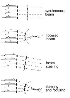
Probes are usually constructed of a collection of piezo crystals placed side by side, along the length of the transducer head (Figure 1). Each generates a pulse of sound radiating out in all directions. If all are stimulated at the same time, then constructive interference generates a wave-front parallel to the head. If, however, each crystal is stimulated after a brief delay, then the effect is to angle the wave-front.
Phased Array
Doppler effect
Principle and clinical applicationDoppler ultrasound
- Uses transducer crystals to transmit and receive ultrasound
- Transducers may be placed directly on the skin
- Can be used to measure blood pressure
- Is unaffected by movement
- Measurements are affected by diathermy
TFTFT